Can Dogs Get Concussions? How to Identify & Treatment
What are concussions?
Table of Contents
A concussion is a condition that is caused by a head injury. Various reasons can cause the damage i.e. hit, jolt or bump on the head. Most concussions in dogs are caused by car accidents, falling and severe fights with other dogs/animals. It can also be caused by a hit on the body that further causes the brain to move back and forth abnormally. The irregular movement caused by the injury creates chemical changes in the brain due to the movement and can damage brain cells.
Can dogs get concussions?
Yes, dogs can get concussions and it’s very common head trauma in dogs. Most outdoor dogs are at more risk of getting a concussion due to the traffic on roads. Dogs often get hit by vehicles and that causes head injury in them. There are various other reasons why your dog can get a concussion. Any dog that is harshly hit by anything can be a victim of traumas. It does not mean your pet dog is not at risk, they must be taken care of and their surroundings must be safe.
Symptoms of concussions in dogs:
Now that you know dogs can get concussions, how to identify them is another thing to learn.
Whenever we humans have any medical issue, we can express it and talk about it. But, when dogs or other animals have any health issues, they cannot verbally express but they try to convey their pain and discomfort through other signs which must be paid attention to.
If the dog is not experiencing pain after being hit or getting a jolt, it gets hard to identify concussions. But, there are a few expected symptoms that your dog will experience as listed below:
There are various symptoms they experience when dogs get a concussion. It also depends on the kind of injury or accident that they have been through.
Some dogs go through seizures after injury.
- Abnormal eye movement.
- Vomiting can be caused after the accident.
- Bleeding.
- Body paralysis makes them unable to stand or move.
- The size of their pupils might change.
- Dullness
- Lethargy
- Fainting
- Breathlessness
If any of these symptoms appear after an injury, do not ignore the signs and inform a vet right away.
Treatment for concussions in dogs:
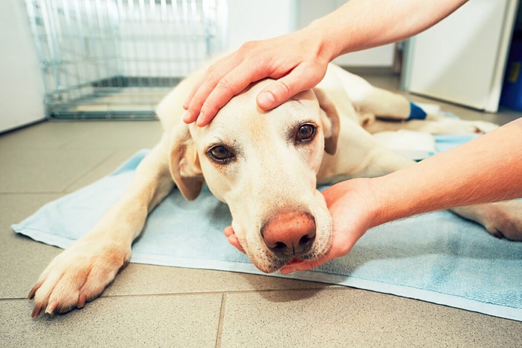
The right thing to do after your dogs have been hit or had an accident is to call their vet and inform them of the accident, if required, take them to the nearest veteran right away (in case of a car accident or other major hit). While you need to rush, do not panic and do not be harsh on the dog. Any wrong position or movement can worsen the situation. It is suggested to keep the head elevated by keeping some support so that minimum pressure must be on the brain. If the dog cannot walk on its own, it might need a stretcher and you need to call an ambulance right away in that case.
What happens next?
The veteran will start the treatment process according to the severity of the injury. They will initially check all the vitals and blood pressure to ensure that the dog is stable to go through the next treatment processes. The vet will later do some neuro tests to check if there is any internal bleeding or swelling of the brain. The later treatment will be done according to reports of the tests. If the vet keeps your dog on oxygen support, there is nothing to worry about as this will be a prudent step to prevent any problem. The later treatment will depend on the situation of the brain and will only be advised and monitored by the vet.
In most cases, there is no special treatment given in mild cases of concussion in dogs. Instead, they are given the right care and time to heal themselves. But, in severe cases, the treatment will vary accordingly. As the dog owner, you will have to put in a lot of time and concentration to make sure the healing is on the right track and the situation is not worsening.
You will need to give your dog a break. They need to rest and all the play activities and walk routines can wait for a while because you want to prevent any discomfort in the way of their healing. If you take them out, they may hurt themselves again, which will worsen the situation. At home, make sure there is nothing in the dog’s proximity to get hit by.
If there is an external wound, the vet will instruct you for dressing and some medication or creams to be applied.
A good diet and intake of all the essential nutrients are very important for healing. You must make sure that they are eating enough and healthy. If they do not eat, the healing process will become very slow and this can also cause some permanent damage. The healing process is a critical time and you need to be with them through it. They will need constant attention and observation. If they are taken care of, they will heal fast but if you notice they’re not healing and instead of showing more symptoms, you must inform the vet and seek their advice on the matter.
Are concussions in dogs serious?
Usually, concussions can be treated if medical help is sought urgently. But, if the treatment is delayed, this can cause permanent brain damage.
To prevent any risk factors, medical help must be sought urgently after any symptoms have been observed.
In conclusion, as a human, you need to take care of your pet dogs and the stray dogs on the streets. Do not drive fast and be mindful and careful of the living beings around you. Your one mistake can take away someone’s life. If any dog is hit by your car or if you see a dog or any other animal lying helplessly somewhere, it is your responsibility to provide help.
Causes and varieties
There are many reasons why a concussion in a dog can occur. In most cases, they are similar to accidents that cause the same consequences in people:
Traffic accidents. This is the most common cause – dogs often get hit by cars and suffer head injuries of varying degrees of intensity.
Falls. A dog can be injured even if it falls from a small height. Smaller members of the dog population can be injured by falling out of the owner’s hands or out of a special bag or carrier.
Head injuries associated with blows to the skull. It can be careless actions of the animal itself or the owner who dropped something heavy on the dog, as well as the violent actions of immoral individuals who beat the dog. Falling something heavy on the head can cause not only a concussion, but also various injuries to the skull, up to and including open wounds that require immediate response and immediate medical attention.
Gunshot wounds to the head. Hunting breeds are most often affected, but similar incidents can happen to other animals as well. Fortunately, this is a rare case of injury.
Sometimes dogs self-injure, resulting in a concussion in the dog. While hunting, playing, active running, it can hit hard on various objects – furniture corners, wall ledges, trees, large stones and so on. A blow to the head may cause a concussion.
According to the degree of intensity, head injuries that provoke a concussion are divided into:
- Light. They do not threaten the health and life of the dog.
- Medium. They can affect the pet’s well-being and health.
- Severe. Such lesions are life-threatening.
The causes of concussion in dogs are varied, but the consequences in all of them can be very dangerous.
Therefore, such conditions require careful attention and quality treatment, taking into account the severity of the lesion, weight, size and age of the animal.
First Signs
A dog that has suffered a concussion may behave as normal with a mild injury. With more severe lesions, the following signs and symptoms of a dog concussion occur:
- Drowsiness, inert behavior, and apathy. These are usually the first signs of a mild concussion.
- Headache, which causes the animal to either try to keep the head unnaturally straight and still, or shake it vigorously.
- Movement coordination disorders.
- Nausea, vomiting.
- Lack of appetite.
- The dog sleeps a lot, tries to hide from everyone, even from the owners.
- Loss of consciousness, sudden fainting. Loss of consciousness is especially dangerous if the animal is constantly vomiting – it can choke on its own vomit.
- Inadequate behavior up to attacking its owner and bystanders.
- In severe cases, breathing and heartbeat disorders occur, and the pupils are dilated across the eye and unresponsive.
Depending on the condition, the dog is treated accordingly.
First Aid
If a dog has suffered a concussion, it is absolutely necessary to help him. To do this, the following steps are performed:
- You need to remove the dog’s collar, muzzle, anything that may constrain its movements and breathing.
- Lay her on her side on a flat hard surface.
- Pull out his tongue so he cannot choke or suffocate if he loses consciousness.
- Check her pulse and breathing.
- Put a cold compress on the head (ice pack available).
- Seek veterinary help.
If your dog has been hit by a car or has additional injuries, especially fractures and open wounds, you should try to call an emergency vet or house doctor.
Making a diagnosis at the vet clinic
At the hospital, specialists will immediately notice that the pet’s pupils are dilated over the entire eye and send it for a CT scan. With this modern diagnostic method, the level of brain damage and its area of spread can be quickly determined.
Therapy and Treatment
Treatment of a concussion in a dog should only be professional. Self-treatment can lead to unpredictable consequences up to the death of the dog. Only a veterinarian can treat such a dangerous lesion. He will determine the condition of his patient and choose individual treatment techniques based on the level of damage, age, weight and breed of the dog.
In the event that a head injury causes impaired consciousness, if the dog is unresponsive to external stimuli, you need to contact the veterinarian immediately.
Delay can be extremely dangerous. Medics prescribe treatments that are very similar to those for concussions in humans, but with the correction that the medications must be for veterinary purposes.
Self-treatment is extremely dangerous, so any medication should only be used if the veterinarian decides and judging the dog’s condition. It must be remembered that some “human” medications can lead to extremely undesirable consequences.
If the pet has suffered a head injury, the first thing to do for him at home is to ensure complete rest, warmth, no drafts, plenty of clean water, light nutrition. If the animal’s condition does not improve for several days, it should be taken to a clinic.
Pet owners are concerned about how long a concussion heals. This depends on the severity of the injury. Mild injuries will take anywhere from a few days to a couple of weeks, concussions of moderate severity need a longer recovery period, and severe injuries take months to rehabilitate.
Consequences
The consequences of the condition depend on the extent of the lesion and the severity of the injury. If the level of nerve damage is severe, there may be impaired mobility, paresis and paralysis, impaired coordination of movement, vision and hearing, intestinal atony, and other dangerous problems.
With mild damage, a traumatized animal can fully recover without any effects on the body or its functioning.
Prevention
In order to ensure that the dog is not injured, his owner should take care to do the following:
- Take her out for a walk on a leash and keep her off the leash in dangerous places.
- Remove any potentially dangerous items from the house and yard.
- Do not allow children to torture or terrorize your pet.
- Take your pet to the veterinarian for regular checkups.
Conservative treatment of dogs with craniocerebral trauma
Lethality in severe traumatic brain injury remains very high – 60-80%.
A distinction is made between primary and secondary brain injuries. Primary injuries include those that are a direct consequence of trauma: skull fractures, ruptures of blood vessels, and rupture or crushing of the brain parenchyma. Secondary injuries develop due to anatomical and physiological changes: brain edema, hematomas, increased intracranial pressure, ischemia. These changes occur within hours or days after injury, and their correction is the primary task of the ORIT physician.
Initial measures

in case of vital organs dysfunction, the examination must be preceded by emergency measures – tracheal intubation, ventilatory ventilation, BP normalization;
determination of the depth of impairment of consciousness (before consulting a neurologist), for which the Glasgow Coma Scale, adapted for animals, is most often used:
| Motor activity | Normal gait, normal reflexes Hemiparesis, tetraparesis, decerebral rigidity Position with intermittent extensor stiffness Position with permanent extensor stiffness Position with intermittent extensor stiffness/opistotonus Hypotonic position with depressed/absent spinal reflexes |
6 5 4 3 2 1 |
| Stem reflexes | Normal oculocephalic and pupillary reflexes Slow pupillary reflex and normal or impaired oculocephalic reflex Bilateral/permanent miosis and normal or impaired oculocephalic reflex Pinpoint pupils and impaired or absent impaired oculocephalic reflex Unilateral mydriasis and impaired or absent oculocephalic reflex Bilateral/permanent mydriasis (“doll’s eyes”) and impaired or absent oculocephalic reflex |
6 5 4 3 2 1 |
| Level of consciousness | Normal Depressed/delirious, but able to respond to stimuli Inhibited/stupor but able to respond to visual stimuli Inhibited/stupor but able to respond to acoustic stimuli Inhibited/stupor but able to respond to painful stimuli Comatose but unable to respond to painful stimuli |
6 5 4 3 2 1 |
| Overall estimate of the prognosis | 3-8 unfavorable 9-14 cautious 14-18 good |
IMPORTANT: A complete neurological examination should be repeated after 30-60 minutes, then after 3 hours, and then daily, depending on the severity of the injury and the initial examination. The course of therapy should be changed if there are changes in the nervous system.
- MRI, CT scan (if available);
X-ray of the chest, skull in two projections, cervical spine;
IMPORTANT: One in ten patients with severe traumatic brain injury has a cervical spine injury at the same time. As long as spinal injury has not been ruled out, the animal must be treated as having a spinal cord injury.
Laboratory tests: total blood count, blood glucose, blood gases and electrolytes, blood creatinine and urea, and total urine analysis;
monitoring: general clinical – BP, HR, BP, ECG, blood saturation; neuromonitoring – IOP, EEG, transcranial Doppler echography.
The construction of treatment tactics depends on the specific clinical situation:
- type of injury: epidural hematoma, subdural hematoma, brain contusion, depressed skull fracture, combination of these conditions;
- volume of injury;
- degree of consciousness deprivation (DCI);
- results of monitoring.
Treatment of animals in the acute period of traumatic brain injury
Priority measures should be completed within 2 hours of the animal’s admission to the ORIT.
If the animal has aspiration syndrome and disturbed consciousness (coma), immediate tracheal intubation and sanation of tracheobronchial tree should be performed. Orotracheal intubation is preferable.
Emergency therapeutic and diagnostic bronchoscopy is indicated in the presence of solid food particles in aspirated fluid, progression of acute respiratory failure.
Placement on the ventilator
Indications for artificial ventilation are coma, hyper- and hypoventilation syndrome, respiratory disturbance, signs of increasing intracranial hypertension, associated thoracic injuries, traumatic shock of degree 2-3, signs of decompensated respiratory failure of any genesis. Positive end-expiratory pressure (PEEP) of 2-4 cm a.c. should be used whenever possible in all animals with TMT, since it reduces atelectasis formation and does not increase IAP. Synchronization of the animal with the ventilator using sedatives or non-depolarizing muscle relaxants is mandatory.
IMPORTANT: If there is no possibility of ventilating the animal, it is worth refraining from administering narcotic analgesics to the animal.
Stabilization of hemodynamics
It should always be assumed that traumatic brain injury develops cerebral ischemia, so blood volume should be restored promptly to improve blood flow to the brain. It is extremely important to maximize oxygen supply and minimize the risk of excessive fluid intake. For this purpose it is best to monitor CVP and BP, acid-base balance of blood, body temperature, HR and heart rate (ECG), respiratory rate and rhythm, diuresis, and use oxyhemometry. Measures should be taken to restore normovolemia. Routine use of diuretics is unacceptable, since it may adversely affect the outcome of the disease. The best method, recognized in many countries of the world, is a combination of infusions of colloids and hyperosmolar solutions. This method is called the low-volume resuscitation method. For example, infusion of 7.5% sodium chloride combined with colloid (6%) quickly restores CPR and does not lead to IOP increase. If there is no possibility to use sodium chloride, or there are electrolyte disorders (hypernatremia, hyperchloremia), it is necessary to use another hyperosmolar solution – mannitol 15%.
IMPORTANT: Mannitol actively removes K from the body. This should be monitored daily until this solution is withdrawn. Mannitol is administered in a dose of 1.5 g/kg every 6 hours in the first day, with a dose reduction to 0.5 g/kg in subsequent days.
Elimination of hypoxia
Optimal PaO2 level is 80-90 mmHg. Additional oxygen will not harm animals with CHT, but it must be remembered that blood oxygen levels are also directly related to red blood cell counts. Anemia must be eliminated. It is also important to remember the benefits of normoventilation and try to maintain it.
IMPORTANT: At low BP, in conditions of eliminated hypovolemia, in the absence of pneumothorax and hypoxia, dopamine should not be used as a vasopressor, as it is more likely to increase cerebral edema. If BP is elevated, hypotensive therapy may be delayed, as BP will most often normalize with the start of sedation therapy.
The animal should also be given an elevated head position of at least 10 and no more than 30 degrees, as this position produces maximum gravitational IOP reduction without significantly affecting cerebral perfusion pressure or cerebral blood flow.
Sedation Therapy
Adequate sedation therapy reduces the metabolic demands of the brain, reduces the likelihood and progression of intracranial hypertension, and facilitates synchronization of the animal with the ventilator. Sedation therapy is necessary during the acute period. The criteria for its adequacy include: absence of motor agitation, absence of tachycardia, preservation of moderate spontaneous muscle activity (exception – animals with HFH), cough reflex, reaction to painful stimuli. In deep coma and good synchronization with the ventilator, sedatives are either not used or used in minimal dosages. In animals with HFH, on the contrary, sedation should be deeper. If the animal is on spontaneous breathing and the degree of trauma is low, sedation therapy consists of maintaining a drowsy state (without the use of narcotic analgesics).
Continuous infusion provides greater hemodynamic stability. Propofol or sodium thiopental are used. For unstable hemodynamics, it is better to use phenazepam also as a continuous infusion.
Correction of intracranial hypertension (ICH)
Unfortunately, it is not possible to adequately assess the presence and degree of intracranial hypertension, since there are no methods to measure intracranial pressure in animals. We can indirectly judge about intracranial hypertension by such symptoms as behavioral disorders (hyperactivity), vision disorders, changes in ocular vascular pattern, changes in heart rhythm (bradycardia), ventricular dilation on neurosonogram, etc. The correction of HFH consists mainly in its prevention.
Prevention of early seizure syndrome.
The drug of choice is phenobarbital. Seizure syndrome prophylaxis has a tangible effect on sedation doses, significantly reducing them and reducing the risk of cardiovascular and respiratory system disorders. Seizures are also an independent risk factor for animal death and should be eliminated.
Neuroprotection
Taking into account the fact that several cascades of reactions causing cell death are activated during a CHT, effective neuroprotection requires a complex combination of drugs acting on different links in the chain of pathophysiological events as the brain damage evolves. The most effective drugs are erythropoietin, progesterone, and cyclosporine. Their use during the acute period of injury has been shown to significantly improve outcome.
IMPORTANT: The therapeutic “window” for these drugs is limited to 6-12 hours after injury. Their use outside this range significantly reduces their effectiveness.
Feeding
On the second day after admission of an animal with a traumatic brain injury to the ORIT, enteral (probe) feeding should be started. Improvement in prognosis has been proven in 3 randomized study groups of animals. The caloric content of nutritional mixtures should be increased, starting with a hypocaloric diet and gradually bringing it up to the normal needs of the animal. Enteral nutrition is replaced by parenteral nutrition if the gastrointestinal tract cannot be used.
IMPORTANT: Blood glucose levels may be elevated in CHF. Hyperglycemia increases anaerobic metabolism and may lead to cerebral acidosis. Monitoring should be done every 4-6 hours, and the rate of nutritional supplementation is adjusted according to the results, and insulin is administered if necessary.
Care
To prevent bedsores and pulmonary atelectasis, the animal should be turned over every 4 hours. Physical therapy is desirable to prevent muscle contractures.
IMPORTANT: Patients with multiple musculoskeletal injuries or skull fractures experience pain, so adequate analgesia must be provided.


This insightful article provides a comprehensive look into the often misunderstood and overlooked topic of canine concussions. It underscores the importance of dog owners recognizing the signs and symptoms of concussions in their pets, including one symptom in particular – drowsiness.
It’s remarkable how this subtle sign can often be misinterpreted or entirely missed, with owners attributing it to mere tiredness after play. The feature does a commendable job of highlighting the serious implications behind such signs, particularly when they occur in connection with an incident like a fall.
The story of the dog who had recently fallen off a small cliff stands out. It’s an eye-opener, as it allows readers to grasp the significance of vigilance and prompt medical attention in such scenarios. By recognizing the abnormal drowsiness in their pet, the owners were able to act swiftly, potentially saving their beloved dog from severe neurological complications.
This article serves as a stark reminder that our furry friends cannot communicate their discomfort in the same way humans can. It’s up to us to stay informed, notice the signs, and ensure their safety and well-being.
On behalf of the reader, I want to extend a heartfelt thank you for this invaluable information. By delving into the complexities of canine concussions and explaining their symptoms clearly, the article has undoubtedly empowered many dog owners, ensuring their pets receive the best possible care in the event of an unfortunate accident.
In summary, this piece is not just an article but a guide, a tool for pet owners to keep their companions safe. As a society, we are becoming more aware of the importance of mental health, and this should extend to our pets as well. I urge all pet owners and lovers to take the time to understand these crucial signs.